The Delicate Art of Treating Festoons: Why the Wrong Treatment Makes Them Worse
Festoons, the soft, hammock-like pouches that drape across the upper cheek below the lower eyelid, are one of the most humbling conditions in periorbital aesthetics. They are progressive, they are multifactorial, and they respond unpredictably to treatments that work beautifully elsewhere on the face.
This is a condition we discuss extensively and teach to our fellows: physicians who come to train with us after they have already become cosmetic specialists. It demands that level of nuance. Getting it wrong does not just fail to improve the problem. It can make it visibly worse.
What exactly are festoons?
Festoons are chronic collections of edematous soft tissue in the infraorbital and malar regions. Unlike simple under-eye bags (which are herniated orbital fat), festoons involve a complex interplay of weakened ligaments, impaired lymphatic drainage, and progressive tissue laxity.
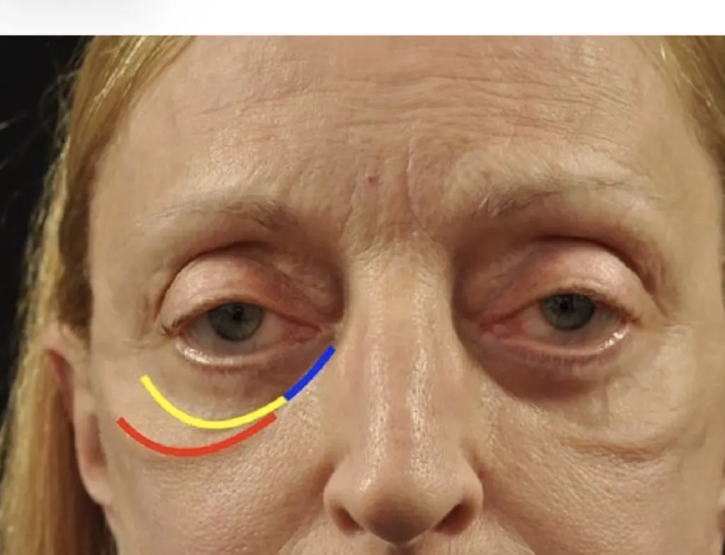
The anatomy is intricate. Three key structures define the festoon boundary: the orbicularis retaining ligament, the maxillary attachment of the orbicularis muscle to the orbital rim, and the zygomaticocutaneous ligament. When these weaken, fluid accumulates and the tissue sags, creating the characteristic mound.
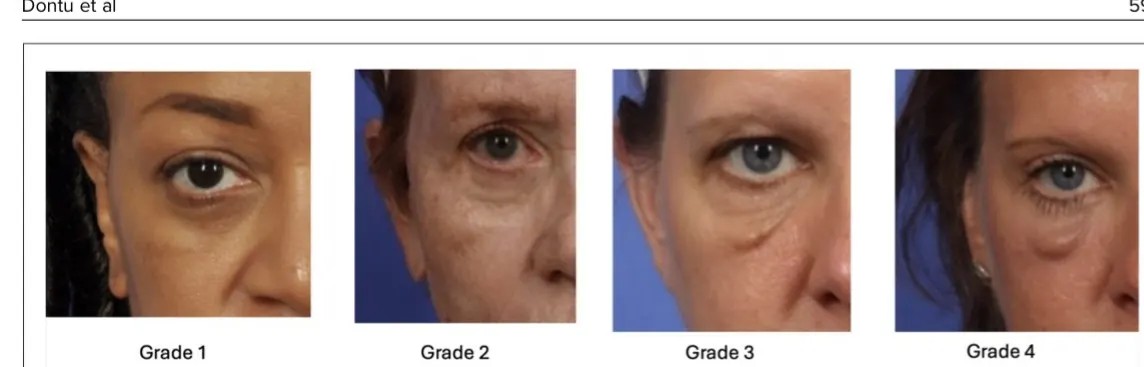
Critically, festoons are progressive. What starts as a Grade 1 delineation line can advance to a Grade 3 or 4 mound if left unaddressed or treated incorrectly. Early intervention is not optional: it is the difference between a manageable condition and one that requires complex surgical correction.
The festoon grading scale
Festoons are classified on a 0 to 4 scale. Catching them at Grade 1 or 2 gives us the best chance of meaningful improvement with non-invasive approaches. By Grade 3 or 4, the tissue changes are more established and treatment becomes significantly more complex.
| Grade | Presentation |
|---|---|
| Grade 1 | Subtle line at the inferior orbicularis |
| Grade 2 | Visible delineation defining the mound |
| Grade 3 | Moderate mound with tissue redundancy |
| Grade 4 | Severe protrusion with significant laxity |
Book RF microneedling at deRMA Skin Institute. Assessed and treated by Dr. Sajic in Guelph. Request a consultation to be graded first. Book now →
Time is tissue: why catching festoons early changes everything
Progressive by nature. Festoons do not stabilize on their own. The combination of UV damage, gravity, and impaired lymphatic drainage means they worsen year over year. What you see today is the mildest it will ever be.
The treatment window narrows. At Grade 1 or 2, radiofrequency microneedling and conservative approaches can produce meaningful improvement. By Grade 3 or 4, the tissue changes are structural, and often require surgical intervention.
Wrong treatment makes it worse. Fillers injected in the wrong plane, excessive volumes, or poorly planned interventions can worsen festoons by creating chronic edema. Early, correct treatment avoids this trap entirely.
How the wrong approach makes festoons worse
This is what makes festoons so treacherous: treatments that work beautifully for other periorbital concerns can actively worsen malar mounds. The tissue here is uniquely unforgiving.
- Fillers in the wrong plane. Hyaluronic acid fillers can block lymphatic drainage, creating chronic edema that inflates the festoon rather than camouflaging it.
- Wrong energy depth. RF microneedling at incorrect depth settings fails to reach the target tissue, or worse, creates inflammation that swells the mound.
- An aggressive single-session approach. Attempting to resolve festoons in one aggressive treatment often triggers a rebound inflammatory response that makes the condition more visible.
- Ignoring the sequential protocol. Festoons require treatments to be layered in a specific order. Skipping steps or combining modalities incorrectly compounds the problem.
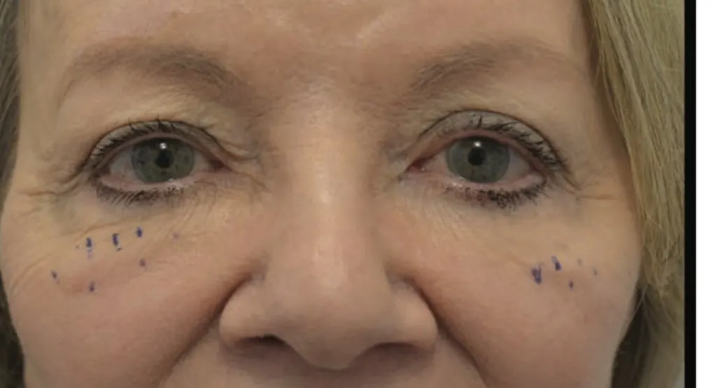
Had a treatment that made things worse? A second opinion from a fellowship-trained specialist can help. Precise injection mapping matters here: each mark on the skin represents a calculated decision about depth, volume, and tissue plane.
The correct protocol: sequential, preferential pairing
Festoons cannot be solved with a single modality. The condition demands a layered, sequential approach where each treatment is chosen based on the response to the previous one. This is what we mean by "preferential pairing": the next step is always determined by how the tissue responds, not by a fixed menu.
- Assessment and grading. Clinical grading, anatomy mapping, and lymphatic function evaluation. No treatment begins without this.
- Foundation treatment. RF microneedling at calibrated depth (2.0 to 2.5 mm) to initiate collagen remodeling without inflammatory rebound.
- Response evaluation. A reassessment at 4 to 6 weeks. The tissue tells us what it needs next, not a predetermined protocol.
- Layered refinement. Subsequent sessions adjusted in depth, energy, and modality based on tissue response. This may include sclerotherapy or fat repositioning.
The depth question
In the largest study to date on RF microneedling for festoons (Dontu et al., 2025; 43 patients), treatments used calibrated passes at 2.5 mm, 2.0 mm, and 1.5 mm depths with precise millisecond energy delivery. The mean festoon grade showed statistically significant improvement, but only when depth was matched to tissue thickness. Too shallow: no effect. Too deep: inflammation and worsening. This is why festoon treatment is a fellowship-level skill.
What correct treatment looks like
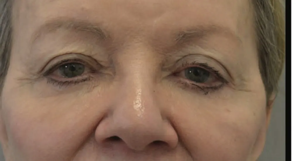
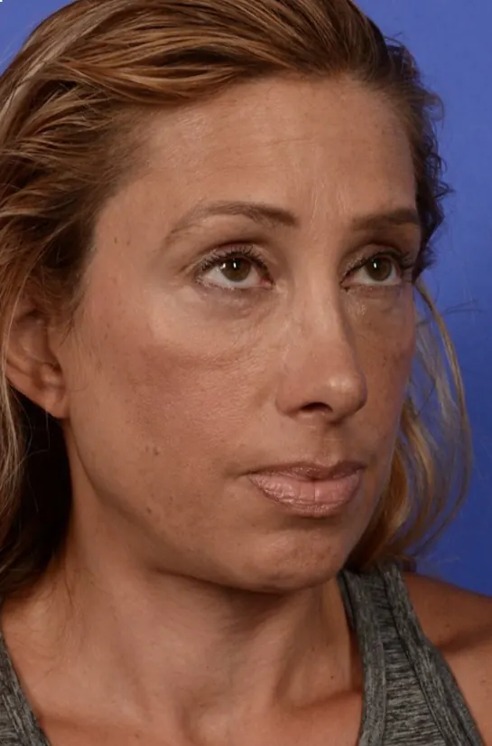
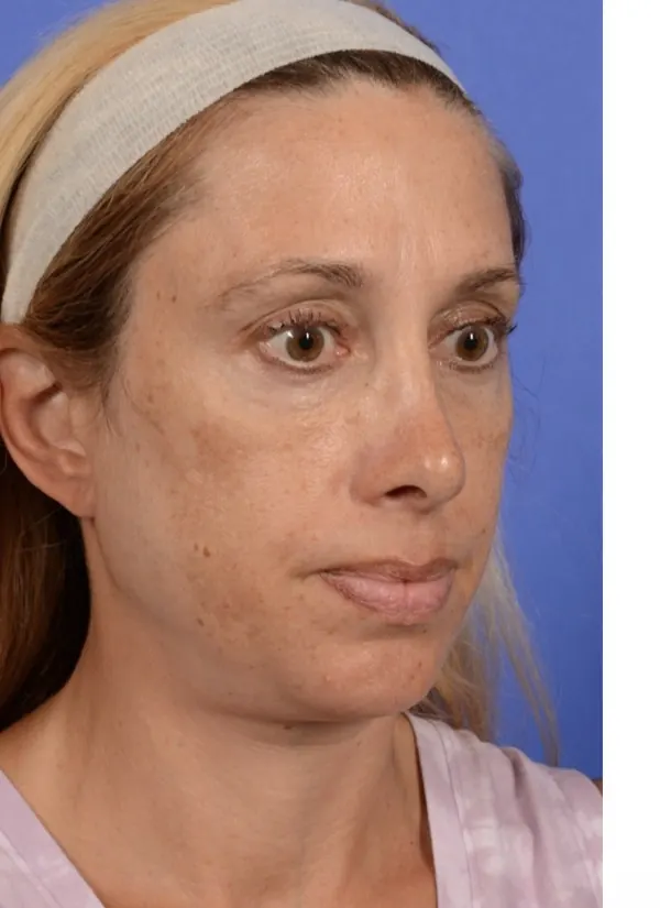
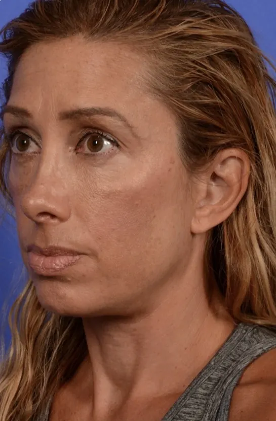
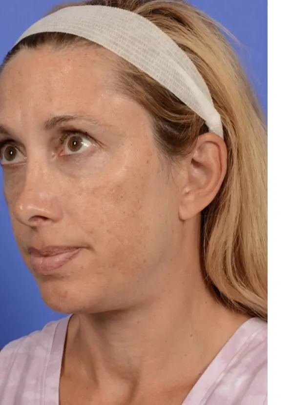
Published and clinical results demonstrate what is achievable when the right professional applies the right protocol at the right depth: sequentially, over multiple sessions, with careful reassessment between each one. In treated cases we see reduction in malar mound volume, improved skin quality, and consistent bilateral improvement achieved through sequential RF microneedling. Clinical photographs of these outcomes are used with permission for educational purposes, and individual results vary.
Why this is a fellowship-level skill
Festoons are one of the conditions we teach extensively to our fellows, physicians who come to train with us after they have already completed their specialty training and become cosmetic specialists. This is not a beginner procedure. It requires:
- Deep understanding of periorbital ligament anatomy and tissue planes
- The ability to grade festoons accurately and match treatment intensity to grade
- Experience calibrating RF microneedling depth to individual tissue thickness
- Judgment to know when to stop, when to wait, and when to change approach
- Recognition of when surgical intervention is the honest recommendation
The benefit of seeking the right professional far outweighs the cost of convenience. A single poorly executed treatment can set you back months or years. The right specialist will give you an honest assessment, even if that means telling you what you do not want to hear. Our fellowship program trains post-residency physicians in advanced periorbital techniques, including the nuanced, sequential approach to festoon management that cannot be learned from a weekend course. You can read more about Dr. Sajic and the training program here.
Frequently asked questions
What is the difference between festoons and under-eye bags?
Under-eye bags are typically caused by herniated orbital fat pushing forward through a weakened orbital septum; they sit directly below the lower eyelid. Festoons (malar mounds) are different: they are chronic collections of edematous tissue that drape across the upper cheek, below the orbital rim. They involve weakened ligaments, impaired lymphatic drainage, and progressive tissue laxity. The distinction matters because treatments that work for bags can worsen festoons.
Can fillers fix festoons?
In most cases, fillers are not recommended for festoons and can make them worse. Hyaluronic acid fillers can block lymphatic drainage in the malar region, creating chronic edema that inflates the festoon rather than camouflaging it. In very specific cases, fat repositioning (a surgical technique) can camouflage festoons, but this requires deep subperiosteal pockets and is a surgical procedure, not a simple filler injection.
How does RF microneedling help festoons?
Radiofrequency microneedling delivers controlled thermal energy at precise depths to stimulate collagen remodeling and tissue tightening in the malar region. The largest study to date (Dontu et al., 2025; 43 patients) showed statistically significant improvement in festoon grade, with 41 to 63 percent of patients showing improvement in under-eye skin quality. However, the depth settings are critical: too shallow has no effect, too deep causes inflammation. This is why it requires fellowship-level expertise.
Why can't festoons be treated in a single session?
Festoons involve complex tissue changes: weakened ligaments, impaired lymphatics, and chronic edema. Attempting aggressive correction in one session often triggers an inflammatory rebound that worsens the condition. The correct approach is sequential: treat conservatively, wait 4 to 6 weeks, reassess how the tissue responded, then adjust the next treatment accordingly. Each step is determined by the tissue's response, not a fixed protocol.
At what grade should I seek treatment for festoons?
As early as possible. Festoons are progressive; they worsen over time due to UV damage, gravity, and impaired lymphatic drainage. At Grade 1 or 2, non-invasive approaches like RF microneedling can produce meaningful improvement. By Grade 3 or 4, the tissue changes are structural and often require surgical intervention. The earlier you seek assessment, the more conservative and effective the treatment options.
Sources and references
- Dontu P, et al. Treatment of Malar Festoons With Radiofrequency Microneedling. Aesthetic Surgery Journal. 2026;46(6):591–597.
- Geronemus RG, et al. Successful Noninvasive Treatment of Festoons. Plastic and Reconstructive Surgery. 2018;141(6).
- Goldberg RA, et al. Updated Management of Malar Edema, Mounds, and Festoons: A Systematic Review. Aesthetic Surgery Journal. 2020;40(3):246–258.
- Godfrey KJ, et al. Doxycycline Injection for Sclerotherapy of Lower Eyelid Festoons. Ophthalmic Plastic and Reconstructive Surgery. 2019.
- Botti G. Direct Excision of Malar Bags: Back to the Basics. Aesthetic Surgery Journal. 2024.
- Perry JD. How to Improve the Appearance of Festoons (Malar Mounds). Cleveland Clinic ConsultQD. 2025.
- EyeWiki: Festoons. American Academy of Ophthalmology. Updated 2026.
Where this fits at Sajic Longevity. A physician assessment at our Guelph clinic, serving Toronto and the GTA grades your festoons, maps the anatomy, and determines whether conservative or surgical management is the honest recommendation for your specific case. No upsell, just clarity. Book a consultation or call 1-888-430-4665.
Treat festoons in the right order, with the right hands.
Festoons punish guesswork. Sequential RF microneedling by a dermatologist is the approach that helps rather than harms.
The skin longevity method
Fix the core first, then rebuild in sequence.
Read more → Book at the clinicBook RF microneedling at deRMA Skin Institute
Assessed and treated by Dr. Sajic in Guelph. Request a consultation to be graded first.
Book now → Shop Sajic Skin ScienceRejuvenat corrector cream
GMA7 DNA-repair serum for the delicate periorbital skin between treatments.
Shop now →Not sure which is right for you? Call 1-888-430-4665 or email consult@sajicskin.com and our team will point you to the right starting point.
This article is for education and general information only. It is not medical advice, does not establish a physician-patient relationship, and should not replace consultation with a qualified healthcare provider. Individual results vary. Treatment decisions are made only after an individual assessment.